Scientists have made groundbreaking discoveries concerning the formation of human hands and feet, unraveling the intricate processes that oversee their development. Contrary to a simple outward growth, human fingers and toes emerge from within a larger foundational bud. During this process, intervening cells recede, gradually unveiling the distinct digits.
Around the seven-week mark of cell development, a pivotal moment termed “orchestrated cell death” occurs, leading to the revelation of the well-defined shapes of fingers or toes. These insights are part of a comprehensive spatial cell atlas that documents the entire developmental journey of human limbs, providing a remarkable understanding of the underlying mechanisms.
By utilizing special staining techniques, researchers were able to observe how different cell populations strategically arrange themselves, forming discernible patterns as the digits take shape. The unveiling of this spatial cell atlas marks a significant milestone, shedding light on previously unseen processes in the development of human limbs.
This research not only contributes to our fundamental knowledge of limb formation but also holds promise for practical applications. The potential to treat muscle-related disorders or injuries may arise from a deeper understanding of these developmental processes. Moreover, the findings can influence the diagnosis and treatment of congenital limb syndromes, offering valuable insights into the underlying biological mechanisms.
Part of the larger Human Cell Atlas initiative, which aims to comprehensively map every cell type in the human body, this study was conducted by researchers from the Wellcome Sanger Institute, Sun Yat-sen University, EMBL’s European Bioinformatics Institute, and their collaborators. Leveraging cutting-edge single-cell and spatial technologies, the team created an atlas that characterizes the cellular landscape and precisely identifies the location of cells during the early stages of human limb development. Published in Nature, this atlas serves as an openly accessible resource, providing a detailed account of the intricate processes governing limb development in its initial phases.

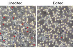
(After two months of development, certain molecules trigger the disappearance of specific cells in the interdigital spaces, revealing the distinct shapes of our fingers and toes.)
The atlas not only reveals novel connections between developmental cells and congenital limb syndromes, such as short fingers and extra digits but also sheds light on the intricate process of limb formation during early human development. Initially, limbs emerge as undifferentiated cell pouches on the sides of the body, lacking a specific shape or function. However, by the eighth week of development, they undergo rapid and precise orchestration, transforming into anatomically complex structures with readily recognizable features like fingers and toes.
The swift and accurate coordination of cells during this process is crucial, and even minor disruptions can lead to downstream effects. Consequently, limb variations rank among the most frequently reported birth syndromes globally, affecting approximately one in 500 births. While extensive studies on limb development have been conducted using mouse and chick models, the degree to which these findings apply to humans remained uncertain. Technological advancements now allow researchers to explore the early stages of human limb formation.
In a recent study, scientists from the Wellcome Sanger Institute, Sun Yat-sen University, and their collaborators delved into tissues between 5 and 9 weeks of development. This timeframe enabled them to track specific gene expression programs activated at distinct times and in specific areas, shaping the evolving limbs. The researchers identified certain gene patterns that play a crucial role in the formation of hands and feet. Disruptions to these genes were linked to specific limb syndromes, such as brachydactyly (short fingers) and polysyndactyly (extra fingers or toes).
Moreover, the study confirmed that many aspects of limb development are shared between humans and mice. The comprehensive characterization of limb development in humans provided by these findings has significant implications. Not only do they offer critical insights into the diagnosis and treatment of congenital limb syndromes, but they also hold potential for addressing muscle-related injuries. Overall, this research contributes to our understanding of the intricacies of limb development, paving the way for advancements in medical interventions and therapies.
“Decades of studying model organisms established the basis for our understanding of vertebrate limb development,” Professor Hongbo Zhang, senior author of the study from Sun Yat-sen University in Guangzhou, China, elaborated. “However, characterizing this in humans has been elusive until now.”
“What we reveal is a highly complex and precisely regulated process. It is like watching a sculptor at work, chiseling away at a block of marble to reveal a masterpiece. In this case, nature is the sculptor, and the result is the incredible complexity of our fingers and toes.”
Dr Sarah Teichmann, senior author of the study from the Wellcome Sanger Institute, and co-founder of the Human Cell Atlas, shared, “For the first time, we have been able to capture the remarkable process of limb development down to single cell resolution in space and time.”
“Our work in the Human Cell Atlas is deepening our understanding of how anatomically complex structures form, helping us uncover the genetic and cellular processes behind healthy human development, with many implications for research and healthcare.”
Recommended For You