Spina bifida is oftentimes a birth defect in which the spinal cord fails to develop or close properly. For unborn babies, this is a devastating diagnosis for the parents who hear this sad piece of news. This has been challenging for doctors, and until recently, these medical experts were unable to attempt to correct the condition while the baby was inside. They had to wait until birth to do anything about it. Even with post-partum medical intervention, the outcome wasn’t always ideal. There were limitations then despite the latest developments of modern technology.
Now, however, discoveries and improvements have been made. Credit should be given to the stunning advances in prenatal surgery because these new operations performed in utero are delivering much more promising outcomes.
Doctors have come up with the theory that the longer spinal tissue is left exposed to amniotic fluid in the womb, the greater the damage to the nerves. The results of which can lead to permanent paralysis of the legs, loss of sensation, and lack of function in the kidney, bladder, and bowels. This can be especially devastating to babies, whose organs have yet to fully develop.
Corrective procedures are oftentimes performed during the second trimester. This means that the operation is done between 23 to 26 weeks of pregnancy. When the intervention happens early on, the medical experts reported that this is able to minimize nerve damage and mitigate long-term health issues. This new procedure may give spina bifida babies the hope of leading lives that are as close to normal as possible.
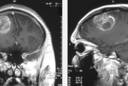
Helena Purcell, an expectant mother from the UK, learned her unborn daughter had spina bifida and hydrocephalus. The latter indicates that there is an abnormal build-up of fluid in the brain. These all happened when she went through her 20-week scan. The doctors saw that half of the baby’s spine was exposed by a large lesion. They had to break the sad news to her and said that the chances her child ever walking were slim. Moreover, her baby would likely be incontinent for her entire life.
The prognosis was bleak and depressing. Within days of receiving this, Helena was tested by the National Health Service (NHS) to see if she qualified for their life-changing in utero surgery program. Fate was on her said because the procedure was approved. Purcell talked to BBC about the whole experience and said, “I knew if I didn’t get the operation the quality of her life would be very different.”
The operation took place when Purcell was 23 weeks into her pregnancy. She flew all the way to Belgium where the surgery had to be done. Almost 30 specialists and clinicians from the University College London Hospitals, Great Ormond Street Hospital for Children, and the University Hospitals Leuven took part in the entire event. They formed a team of fetal and pediatric surgeons, neurosurgeons, anesthesiologists, obstetricians, radiologists, and a scrub team. There were neonatologists on hand as well in case Purcell’s baby needed to be delivered. The neonatologists were there at just the right time because she needed to give birth then.
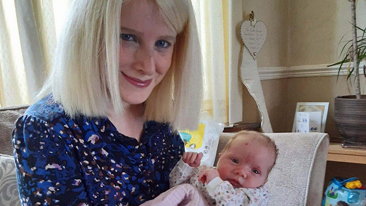
After three months, Helena’s daughter Mila (short for Milagro, which translates to “miracle” in Spanish) was born. She still has some fluid retention in the brain, but her overall development was otherwise good. Purcell also talked to Sky News about this and shared, “I cannot explain the massive difference [this] has had for my family. The NHS doctors are heroes in my eyes, and the surgery they did is just mind-blowing. If it wasn’t for them then Mila would be paralyzed… I am just so grateful that she has had this chance.”
Pre-Born in the USA
The NHS has kept a close eye on this development and reports that since January 2020, 32 British babies and their mothers have undergone the dual surgical procedure. And while many had taken place in the UK, the operation had been successfully performed in the US as well.
This experience also happened to another couple. During a 20-week ultrasound, Mallorie and Chris Deruyter was told that their son, Max, had spina bifida. The Wisconsin couple’s doctors sent Mallorie to Lurie Children’s Hospital in Chicago for further diagnosis and better treatment.
The said operation is known as “closed fetoscopic repair.” This is a much less invasive procedure than the earlier ones. While large leaps have been made, Mallorie still ran a risk for premature delivery. This was a risk that came with the surgery. The doctors made the patients aware that the procedure sometimes induces early birth.
Mallorie shared, “When I initially heard that, I actually thought there’s no way I’m going to have surgery. I just thought it was absolutely crazy. And then the more research I did the more I realized this is going to give him the best life.”
Mallorie went through it and came out of the operating room after a long seven-hour operation. This was helmed by Fetal neurosurgeon Dr. Robin Bowman and pediatric surgeon Dr. Aimen Shaaban. They said that mother and the unborn baby were doing well. The Deruyters went home to Green Bay not too long after and were were set to go back to Lurie for a C-section as soon as the pregnancy reached 39 weeks. Of course, nothing went according to plan. Mallorie went into labor and Max arrived at 3 a.m. just hours before the scheduled C-section. Thankfully, there were no problems.
Baby Max is now finally home and is thriving. The father said, “The chance of a really normal life for him really looks apparent. You can see he’s going to be a thriving, happy young little boy. I don’t think we would have done it any other way.”
3D Printing Brings Forth New Levels of Accuracy
In Florida, on the other hand, along with MRIs and ultrasounds, surgeons are using pioneering 3D printed “virtual” babies. These serve as tools to better guide them through the oftentimes complex steps to perform the procedure.
Orlando Health Winnie Palmer Hospital for Women and Babies in Florida is considered to be one of the best state-of-the-art facilities that makes use of the new technology. Working in conjunction with Orlando-based Digital Anatomy Simulations for Healthcare (DASH), 25 fetal models have since been made. This had been taking place since 2018. Dr. Samer Elbabaa, the medical director of pediatric neurosurgery of Orlando Health spoke and said in a statement, ”The 3D reconstruction of the fetus can really educate the surgeon on the real-life shape, size, and location of the spinal lesion, as well as prepare the surgeon to have the appropriate equipment ready to treat this condition surgically.” He further said, “It’s a level of detail that we are not able to see in traditional imaging, but that is extremely valuable in these cases where we cannot actually see the defect ahead of surgery.”
DASH CEO Jack Stubbs also spoke out about this matter and explained, “The fetal models not only help surgeons plan for things like where to make an incision and how to repair the defect but also help reduce the duration of the surgery to limit the developing baby’s exposure.”
Jocelyn Rodriguez was also a patient at Winnie Palmer. She found out early on that the baby she and her husband Jared were expecting had spina bifida. She was 18 weeks along when they were given the diagnosis. The couple believed that the 3D technology allowed them to better understand what was happening with the pregnancy. Furthermore, they also felt more positive about moving forward with the whole procedure when they had a better picture of what was going to happen.
While Jocelyn hasn’t reached her due date just yet, the subsequent checkups that had taken place after the surgery show how the baby’s condition had already vastly improved with the early intervention. She said, “She has been kicking, wiggling her toes, moving her ankles. She loves to have hiccups. I mean, just everything that we could have wished for has definitely happened.”
Recommended For You